

Introduction #
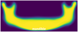
The Panoramic Dental X-rays With Segmented Mandibles dataset provides segmentation of mandibles in panoramic X-rays capturing their anatomical structures. It is composed of almost 2000 digital panoramic X-rays in BMP format of ∼2900×1250 pixels taken by the Soredex CranexD digital panoramic X-ray unit. All the images were taken for diagnostic purposes and treatment planning and no radiography record was taken for the purpose of this study. Two subsets were selected from the original set, but prior to selection, some exclusion criteria were applied to narrow down the sample size. Records with implants were excluded. Also, to ensure that no deciduous tooth was present in the images and that the growth of the jaw had almost completed, patients below 20 years of age were excluded from the dataset. Low-quality X-rays, which were blurred or malposed due to a technician’s error or patient’s lack of cooperation, were also excluded from the dataset.
For the first subset, a maxillofacial radiologist sorted out the panoramic x-rays based on several qualitative features such as the width of ramuses, the vertical distance between the alveolar process and inferior border of the mandible, the acuteness of gonial angle (n), overall convexity of inferior border (g), whether there was a concavity around the gonial angle, shape of coronoid process (m), shape of condyles (p), and depth of sigmoid notch (e). She then purposefully selected 116 images to cover all varieties of mandible shapes seen on panoramic X-rays regarding these criteria. These images were considered templates of the atlas.

For the second subset, 30 images were chosen randomly from the same large dataset and grouped as the statistical set through which the parameters of the system were assigned.
The mandibles of all 146 images were manually segmented by three expert dentists working at Shahid Beheshti University of Medical Sciences, Tehran. In order to generate a reliable unified ground truth for each x-ray, a voting policy was used. According to this voting policy, a pixel belonged to the object (mandible) if at least two of the three manual segmentations defined that pixel as an object:

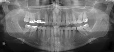
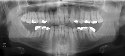
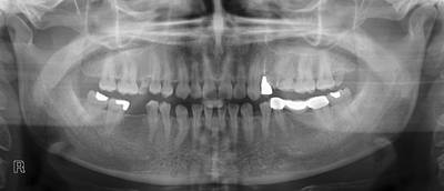
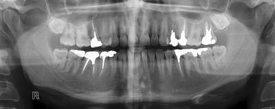
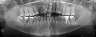
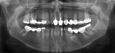
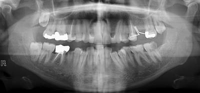
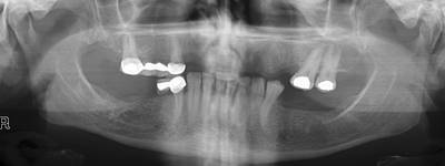
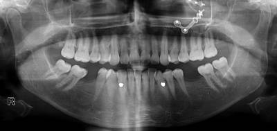
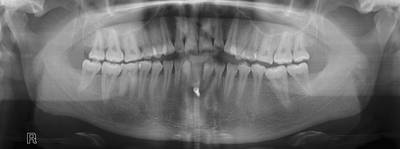
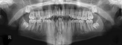
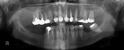
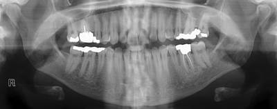
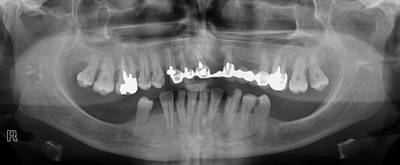
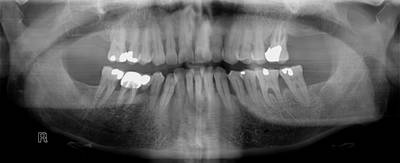
The figure below depicts the outputs of the automatic and manual mandible segmentations in some panoramic X-rays.

The results of the current study seem promising and can lead to further investigations in the field of automatic segmentation of structures in different head and neck radiography techniques. The output of this study can be used in various scenarios, such as improving registrations of panoramic X-rays through the information gained from a segmented mandible. It can affect dental biometrics, which has become a popular field of research. It is also a promising method for human identification.
Intraosseous lesions of the mandible can be detected by performing pattern recognition on the mandible body, which is extracted through the proposed method. Because panoramic radiography is by far the most utilized form of paraclinical record in today’s dentistry (and not all dentists are experienced enough to detect intraosseous lesions), designing a system for autonomous detection of these damaged tissues seems like a good practice that could also result in early detection of lesions. The proposed method can be further extended to detect anatomic landmarks. As such, detecting locations of dental work in panoramic X-rays can be of great use, mostly in cases of dental forensics and detection of periapical lesions.
Please note that on images with index 22 and 110, part of the segmentation is lost and the data was carefully restored by DatasetNinja
 Homepage
 Research Paper
Summary #
Panoramic Dental X-rays With Segmented Mandibles is a dataset for instance segmentation, semantic segmentation, and object detection tasks. It is used in the medical industry.
The dataset consists of 232 images with 232 labeled objects belonging to 1 single class (mandible).
Images in the Panoramic Dental X-rays dataset have pixel-level instance segmentation annotations. Due to the nature of the instance segmentation task, it can be automatically transformed into a semantic segmentation (only one mask for every class) or object detection (bounding boxes for every object) tasks. All images are labeled (i.e. with annotations). There are 2 splits in the dataset: Segmentation1 (116 images) and Segmentation2 (116 images). The dataset was released in 2017 by the Sharif University of Technology, Department of Computer Engineering, Iran and Isfahan University of Medical Sciences, School of Dentistry, Department of Maxillofacial Radiology, Iran.
Explore #
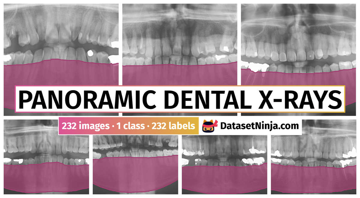
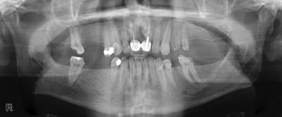
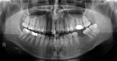
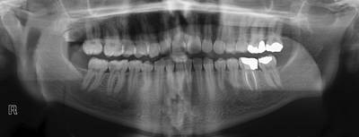
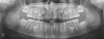
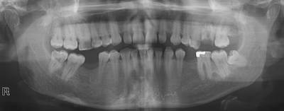
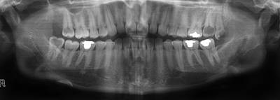
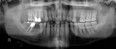
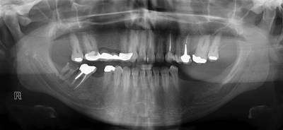
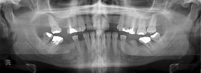
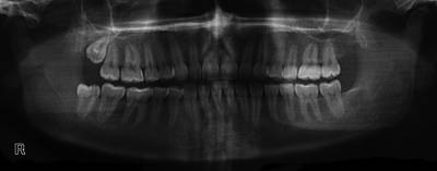
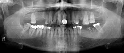
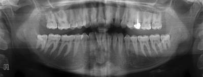
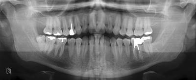
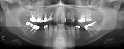
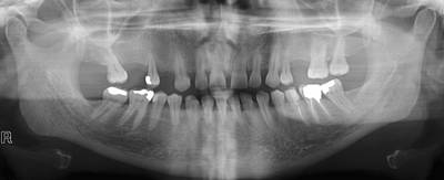
Panoramic Dental X-rays dataset has 232 images. Click on one of the examples below or open "Explore" tool anytime you need to view dataset images with annotations. This tool has extended visualization capabilities like zoom, translation, objects table, custom filters and more. Hover the mouse over the images to hide or show annotations.


Class balance #
There are 1 annotation classes in the dataset. Find the general statistics and balances for every class in the table below. Click any row to preview images that have labels of the selected class. Sort by column to find the most rare or prevalent classes.

Class ㅤ | Images ㅤ | Objects ㅤ | Count on image average | Area on image average |
|---|---|---|---|---|
mandible➔ mask | 232 | 232 | 1 | 39.7% |
Images #
Explore every single image in the dataset with respect to the number of annotations of each class it has. Click a row to preview selected image. Sort by any column to find anomalies and edge cases. Use horizontal scroll if the table has many columns for a large number of classes in the dataset.
Object distribution #
Interactive heatmap chart for every class with object distribution shows how many images are in the dataset with a certain number of objects of a specific class. Users can click cell and see the list of all corresponding images.
Class sizes #
The table below gives various size properties of objects for every class. Click a row to see the image with annotations of the selected class. Sort columns to find classes with the smallest or largest objects or understand the size differences between classes.
Class | Object count | Avg area | Max area | Min area | Min height | Min height | Max height | Max height | Avg height | Avg height | Min width | Min width | Max width | Max width |
|---|---|---|---|---|---|---|---|---|---|---|---|---|---|---|
mandible mask | 232 | 39.7% | 46.65% | 31.36% | 959px | 90.17% | 1330px | 99.76% | 1155px | 95.36% | 2475px | 94.69% | 3117px | 99.9% |
Spatial Heatmap #
The heatmaps below give the spatial distributions of all objects for every class. These visualizations provide insights into the most probable and rare object locations on the image. It helps analyze objects' placements in a dataset.
Objects #
Table contains all 232 objects. Click a row to preview an image with annotations, and use search or pagination to navigate. Sort columns to find outliers in the dataset.
Object ID ㅤ | Class ㅤ | Image name click row to open | Image size height x width | Height ㅤ | Height ㅤ | Width ㅤ | Width ㅤ | Area ㅤ |
|---|---|---|---|---|---|---|---|---|
1➔ | mandible mask | 113.png | 1000 x 2650 | 980px | 98% | 2600px | 98.11% | 40.55% |
2➔ | mandible mask | 2.png | 1330 x 3090 | 1266px | 95.19% | 3084px | 99.81% | 38.95% |
3➔ | mandible mask | 46.png | 1100 x 2760 | 1030px | 93.64% | 2657px | 96.27% | 40.39% |
4➔ | mandible mask | 24.png | 1250 x 2900 | 1195px | 95.6% | 2819px | 97.21% | 39.13% |
5➔ | mandible mask | 79.png | 1280 x 3078 | 1181px | 92.27% | 2997px | 97.37% | 41.96% |
6➔ | mandible mask | 28.png | 1350 x 2700 | 1276px | 94.52% | 2615px | 96.85% | 37.21% |
7➔ | mandible mask | 112.png | 1200 x 2850 | 1103px | 91.92% | 2750px | 96.49% | 34.68% |
8➔ | mandible mask | 23.png | 1245 x 2900 | 1183px | 95.02% | 2832px | 97.66% | 35.51% |
9➔ | mandible mask | 47.png | 1380 x 2800 | 1330px | 96.38% | 2703px | 96.54% | 40.87% |
10➔ | mandible mask | 116.png | 1150 x 2900 | 1118px | 97.22% | 2796px | 96.41% | 41.96% |
License #
Panoramic Dental X-rays With Segmented Mandibles is under CC BY-NC 3.0 license.
Citation #
If you make use of the Panoramic Dental X-rays data, please cite the following reference:
Abdi, A. H., Kasaei, S., & Mehdizadeh, M. (2015).
Automatic segmentation of mandible in panoramic x-ray.
Journal of medical imaging (Bellingham, Wash.), 2(4), 044003.
https://doi.org/10.1117/1.JMI.2.4.044003
If you are happy with Dataset Ninja and use provided visualizations and tools in your work, please cite us:
@misc{ visualization-tools-for-dental-panoramic-x-rays-dataset,
  title = { Visualization Tools for Panoramic Dental X-rays Dataset },
  type = { Computer Vision Tools },
  author = { Dataset Ninja },
  howpublished = { \url{ https://datasetninja.com/dental-panoramic-x-rays } },
  url = { https://datasetninja.com/dental-panoramic-x-rays },
  journal = { Dataset Ninja },
  publisher = { Dataset Ninja },
  year = { 2026 },
  month = { jul },
  note = { visited on 2026-07-15 },
}
Download #
Dataset Panoramic Dental X-rays can be downloaded in Supervisely format:
As an alternative, it can be downloaded with dataset-tools package:
pip install --upgrade dataset-tools
… using following python code:
import dataset_tools as dtools

dtools.download(dataset='Panoramic Dental X-rays', dst_dir='~/dataset-ninja/')
Make sure not to overlook the python code example available on the Supervisely Developer Portal. It will give you a clear idea of how to effortlessly work with the downloaded dataset.
Disclaimer #
Our gal from the legal dep told us we need to post this:
Dataset Ninja provides visualizations and statistics for some datasets that can be found online and can be downloaded by general audience. Dataset Ninja is not a dataset hosting platform and can only be used for informational purposes. The platform does not claim any rights for the original content, including images, videos, annotations and descriptions. Joint publishing is prohibited.
You take full responsibility when you use datasets presented at Dataset Ninja, as well as other information, including visualizations and statistics we provide. You are in charge of compliance with any dataset license and all other permissions. You are required to navigate datasets homepage and make sure that you can use it. In case of any questions, get in touch with us at hello@datasetninja.com.