

Introduction #
Fetal Head UltraSound is a dataset for the measurement of fetal head circumference (HC) - an ultrasound imaging used to measure fetal biometrics during pregnancy. The dataset is a part of HC18 challenge and contains a total of 1334 two-dimensional (2D) ultrasound images of the standard plane that can be used to measure the HC.
All two-dimensional (2D) ultrasound images of the HC were collected from the database of the Department of Obstetrics of the Radboud University Medical Center, Nijmegen, the Netherlands. The ultrasound images were acquired from 551 pregnant women who received a routine ultrasound screening exam between May 2014 and May 2015. Only fetuses that did not exhibit any growth abnormalities were included in this study. Images were acquired by experienced sonographers using either the Voluson E8 or the Voluson 730 ultrasound device (General Electric, Austria). The local ethics committee (CMO Arnhem-Nijmegen) approved the collection and use of this data for this study. Due to the retrospective data collection, informed consent was waived. All data was anonymized according to the tenets of the Declaration of Helsinki.
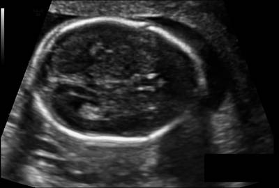
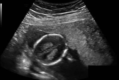
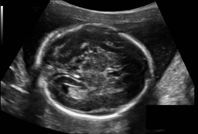
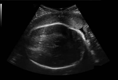
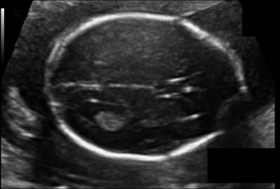
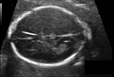
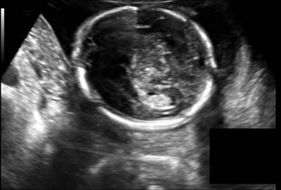
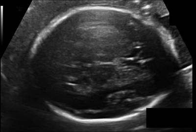
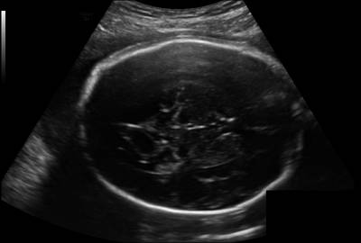
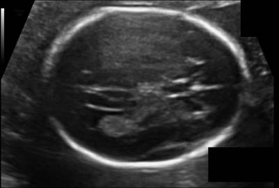
The size of each 2D ultrasound image was 800 by 540 pixels with a pixel size ranging from 0.052 to 0.326 mm. This large variation in pixel size is a result of adjustments in the ultrasound settings by the sonographer (depth settings and amount of zoom are routinely varied during the examination) to account for the different sizes of the fetuses. The size of each 2D ultrasound image was 800 by 540 pixels with a pixel size ranging from 0.052 to 0.326 mm. This large variation in pixel size is a result of adjustments in the ultrasound settings by the sonographer (depth settings and amount of zoom are routinely varied during the examination) to account for the different sizes of the fetuses. Fig. 1 shows example ultrasound images from each trimester. The distribution of the GA in this study is shown in Fig 2. Most data were acquired after 12 and 20 weeks of pregnancy since these are standard time points of routine ultrasound screening for pregnant women in the Netherlands. During each exam, the sonographer manually annotated the HC. This was done by drawing an ellipse that best fits the circumference of the head. Fig. 2 also shows the comparison between the distribution of the HC and the growth curve of Verburg et al.. The reference GA was determined with a CRL measurement between 20 mm (8+4 weeks) and 68 mm (12+6 weeks). All the HCs that fell outside the 3-97 percent confidence interval of the curve of Verburg et al. were individually checked to ensure no mistakes were made during data collection.
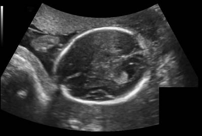
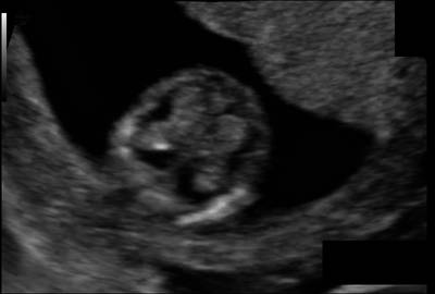
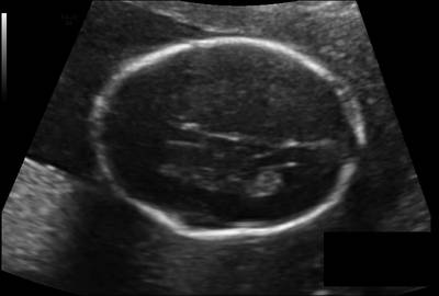
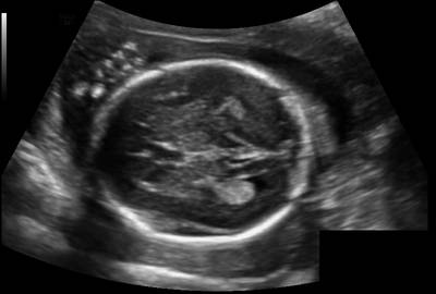
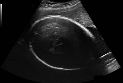
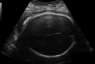
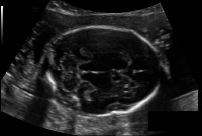
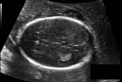
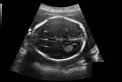
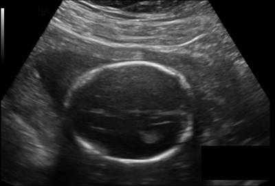
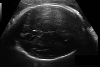
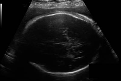
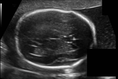
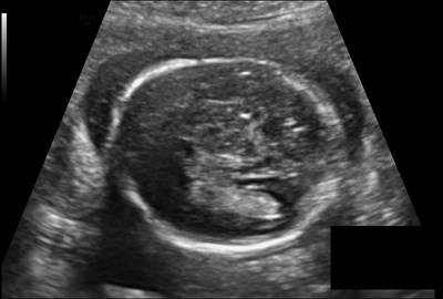
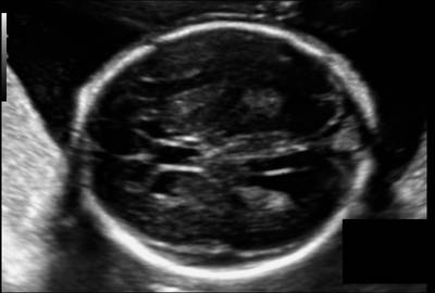
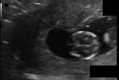
Fig. 1. Example ultrasound images.
Fig. 2. Distribution of HC and GA for the study data.
The data was randomly divided into a train set and a test set of 75 percent (999 images) and 25 percent (335 images), respectively. The GAs were proportionally balanced between the data sets as shown in Table 1. All images that were made during one echographic examination were assigned to either the training or the test set. An independent data set of HC annotations of the images in the test set was created by TLAvdH, a medical researcher who has a technical background in ultrasound imaging and received training from an experienced sonographer in measuring the HC.
| Trimester | Training Set | Testing Set |
|---|---|---|
| First | 165 | 55 |
| Second | 693 | 233 |
| Third | 141 | 47 |
| Total | 999 | 335 |
Table 1. Number of images in the training and the test set.
As a result of HC18 challenge, the following fetal head circumference parameters are calculated:
filename, center_x_mm, center_y_mm, semi_axes_a_mm, semi_axes_b_mm, angle_rad
 Homepage
 Research Paper
 Zenodo
Summary #
Automated Measurement Of Fetal Head Circumference Using 2D UltraSound Images is a dataset for instance segmentation, semantic segmentation, and object detection tasks. It is used in the biological research.
The dataset consists of 1334 images with 999 labeled objects belonging to 1 single class (fetal head circumference).
Images in the Fetal Head UltraSound dataset have pixel-level instance segmentation annotations. Due to the nature of the instance segmentation task, it can be automatically transformed into a semantic segmentation (only one mask for every class) or object detection (bounding boxes for every object) tasks. There are 335 (25% of the total) unlabeled images (i.e. without annotations). There are 2 splits in the dataset: train (999 images) and test (335 images). The dataset was released in 2018 by the Radboud University Nijmegen.
Explore #
Fetal Head UltraSound dataset has 1334 images. Click on one of the examples below or open "Explore" tool anytime you need to view dataset images with annotations. This tool has extended visualization capabilities like zoom, translation, objects table, custom filters and more. Hover the mouse over the images to hide or show annotations.


Class balance #
There are 1 annotation classes in the dataset. Find the general statistics and balances for every class in the table below. Click any row to preview images that have labels of the selected class. Sort by column to find the most rare or prevalent classes.

Class ㅤ | Images ㅤ | Objects ㅤ | Count on image average | Area on image average |
|---|---|---|---|---|
fetal head circumference➔ mask | 999 | 999 | 1 | 30.2% |
Images #
Explore every single image in the dataset with respect to the number of annotations of each class it has. Click a row to preview selected image. Sort by any column to find anomalies and edge cases. Use horizontal scroll if the table has many columns for a large number of classes in the dataset.
Object distribution #
Interactive heatmap chart for every class with object distribution shows how many images are in the dataset with a certain number of objects of a specific class. Users can click cell and see the list of all corresponding images.
Class sizes #
The table below gives various size properties of objects for every class. Click a row to see the image with annotations of the selected class. Sort columns to find classes with the smallest or largest objects or understand the size differences between classes.
Class | Object count | Avg area | Max area | Min area | Min height | Min height | Max height | Max height | Avg height | Avg height | Min width | Min width | Max width | Max width |
|---|---|---|---|---|---|---|---|---|---|---|---|---|---|---|
fetal head circumference mask | 999 | 30.2% | 58.02% | 0.85% | 131px | 24.26% | 505px | 93.17% | 362px | 67.07% | 152px | 19% | 665px | 83.12% |
Spatial Heatmap #
The heatmaps below give the spatial distributions of all objects for every class. These visualizations provide insights into the most probable and rare object locations on the image. It helps analyze objects' placements in a dataset.
Objects #
Table contains all 999 objects. Click a row to preview an image with annotations, and use search or pagination to navigate. Sort columns to find outliers in the dataset.
Object ID ㅤ | Class ㅤ | Image name click row to open | Image size height x width | Height ㅤ | Height ㅤ | Width ㅤ | Width ㅤ | Area ㅤ |
|---|---|---|---|---|---|---|---|---|
1➔ | fetal head circumference mask | 297_HC.png | 540 x 800 | 349px | 64.63% | 432px | 54% | 27.55% |
2➔ | fetal head circumference mask | 874_2HC.png | 540 x 800 | 421px | 77.96% | 484px | 60.5% | 36.69% |
3➔ | fetal head circumference mask | 989_2HC.png | 540 x 800 | 386px | 71.48% | 447px | 55.88% | 31.16% |
4➔ | fetal head circumference mask | 260_HC.png | 544 x 794 | 333px | 61.21% | 455px | 57.3% | 27.59% |
5➔ | fetal head circumference mask | 240_HC.png | 540 x 800 | 245px | 45.37% | 264px | 33% | 11.64% |
6➔ | fetal head circumference mask | 112_HC.png | 540 x 800 | 412px | 76.3% | 437px | 54.62% | 32.15% |
7➔ | fetal head circumference mask | 420_HC.png | 540 x 800 | 385px | 71.3% | 458px | 57.25% | 31.69% |
8➔ | fetal head circumference mask | 214_HC.png | 540 x 800 | 311px | 57.59% | 385px | 48.12% | 21.85% |
9➔ | fetal head circumference mask | 852_HC.png | 540 x 800 | 336px | 62.22% | 395px | 49.38% | 24.13% |
10➔ | fetal head circumference mask | 988_HC.png | 540 x 800 | 416px | 77.04% | 514px | 64.25% | 39% |
License #
Citation #
If you make use of the Fetal Head UltraSound data, please cite the following reference:
- Thomas L. A. van den Heuvel, Dagmar de Bruijn, Chris L. de Korte and
Bram van Ginneken. Automated measurement of fetal head circumference
using 2D ultrasound images. PloS one, 13.8 (2018): e0200412.

- Thomas L. A. van den Heuvel, Dagmar de Bruijn, Chris L. de Korte and
Bram van Ginneken. Automated measurement of fetal head circumference
using 2D ultrasound images [Data set]. Zenodo. http://doi.org/10.5281/
zenodo.1322001
If you are happy with Dataset Ninja and use provided visualizations and tools in your work, please cite us:
@misc{ visualization-tools-for-fetal-head-ultrasound-dataset,
  title = { Visualization Tools for Fetal Head UltraSound Dataset },
  type = { Computer Vision Tools },
  author = { Dataset Ninja },
  howpublished = { \url{ https://datasetninja.com/fetal-head-ultrasound } },
  url = { https://datasetninja.com/fetal-head-ultrasound },
  journal = { Dataset Ninja },
  publisher = { Dataset Ninja },
  year = { 2026 },
  month = { jul },
  note = { visited on 2026-07-14 },
}
Download #
Dataset Fetal Head UltraSound can be downloaded in Supervisely format:
As an alternative, it can be downloaded with dataset-tools package:
pip install --upgrade dataset-tools
… using following python code:
import dataset_tools as dtools

dtools.download(dataset='Fetal Head UltraSound', dst_dir='~/dataset-ninja/')
Make sure not to overlook the python code example available on the Supervisely Developer Portal. It will give you a clear idea of how to effortlessly work with the downloaded dataset.
Disclaimer #
Our gal from the legal dep told us we need to post this:
Dataset Ninja provides visualizations and statistics for some datasets that can be found online and can be downloaded by general audience. Dataset Ninja is not a dataset hosting platform and can only be used for informational purposes. The platform does not claim any rights for the original content, including images, videos, annotations and descriptions. Joint publishing is prohibited.
You take full responsibility when you use datasets presented at Dataset Ninja, as well as other information, including visualizations and statistics we provide. You are in charge of compliance with any dataset license and all other permissions. You are required to navigate datasets homepage and make sure that you can use it. In case of any questions, get in touch with us at hello@datasetninja.com.