

Introduction #
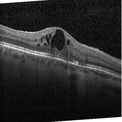
In the study of diversified ocular disorders where cystoid macular edema (CME) is implicated, the authors of the Intraretinal Cystoid Fluid dataset address the significant connection between these disorders and vision impairment. Optical coherence tomography (OCT) scans, used for retina examination, often contain artifacts like blurred edges and speckle noise. These artifacts pose challenges in accurately identifying retinal fluid. The authors employ various image preprocessing techniques, including minimum filtering, block-matching and 3D filtering, and the Richardson–Lucy deconvolution method, to reduce noise and other degradations in OCT scans while maintaining image quality assessment scores. These scores are determined using blind-less image spatial quality evaluation and sharpness estimation.
Furthermore, the authors introduce an automated deep learning (DL) method for detecting and quantifying retinal fluid, specifically CME, to assess its presence and progression. They utilize a U-net model with optimized hyperparameters to achieve optimal results. The improved DL method outperforms previously reported algorithms.
The authors emphasize the potential of combining ophthalmology and artificial intelligence (AI) to revolutionize diagnostic procedures for eye diseases, reduce human errors, and enhance clinical practices. They focus on diabetic macular edema (DME) and its association with cystoid macular edema (CME). They explain the stages of CME development in DME patients and how it can lead to macular holes of varying severity.
The core idea is to use DL algorithms, such as the U-net model, to analyze and predict results from optical coherence tomography (OCT) images, reducing the chances of misdiagnosis. The automated method identifies and quantifies macular fluid in the retina and is compared to expert segmentations to assess its performance.
The authors describe their image preprocessing steps, including resizing, morphological operations, denoising, deconvolution, contrast adjustment, and feature scaling, to enhance OCT image quality and prepare them for DL analysis.
In terms of data collection, the authors obtained a dataset of retinal images primarily focused on DME patients, consisting of 1000 training images and 200 testing images. Manual binary image masking was performed to label pixels as foreground (CME) or background to train the DL model. This dataset was obtained from Kaggle and supplemented with images from the Institute of Ophthalmology.
The study’s ultimate goal is to improve the diagnosis and understanding of CME in DME patients through the application of DL techniques to OCT images, thus reducing the risk of misdiagnosis and aiding medical professionals in their clinical practices.
 Homepage
 Research Paper
Summary #
Intraretinal Cystoid Fluid is a dataset for instance segmentation, semantic segmentation, and object detection tasks. It is used in the medical industry.
The dataset consists of 1460 images with 6542 labeled objects belonging to 1 single class (cystic macular edema).
Images in the Intraretinal Cystoid Fluid dataset have pixel-level instance segmentation annotations. Due to the nature of the instance segmentation task, it can be automatically transformed into a semantic segmentation (only one mask for every class) or object detection (bounding boxes for every object) tasks. All images are labeled (i.e. with annotations). There are no pre-defined train/val/test splits in the dataset. The dataset was released in 2022 by the Mehran University of Engineering and Technology (MUET), Pakistan and Liaquat University of Medical and Health Sciences (LUMHS), Pakistan.
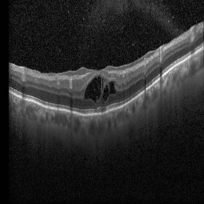
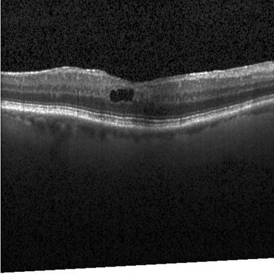
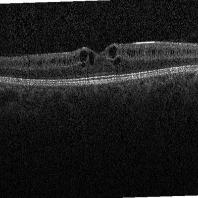
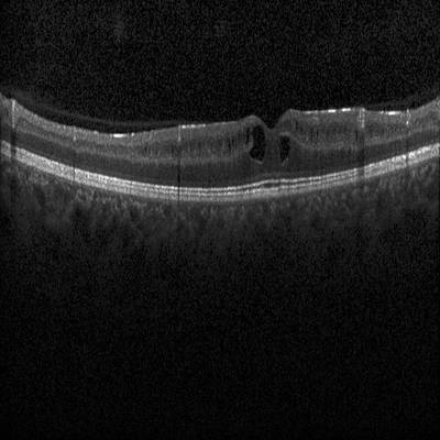
Explore #
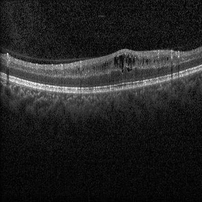
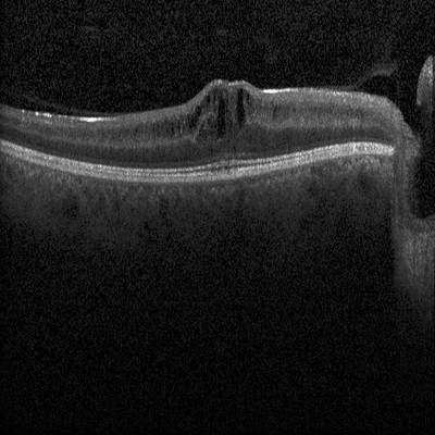
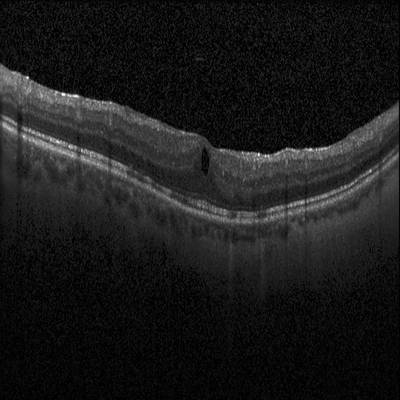
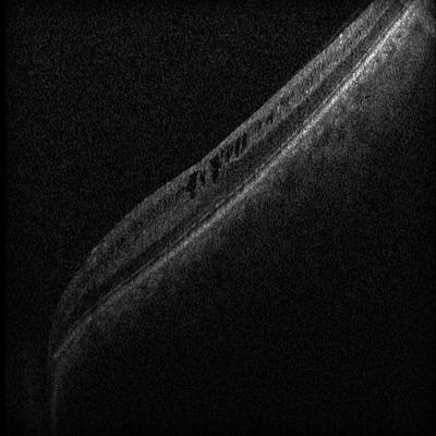
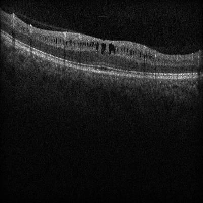
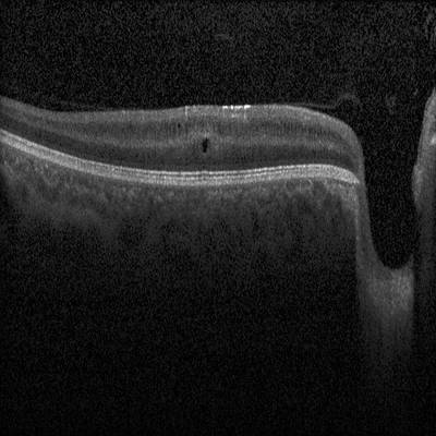
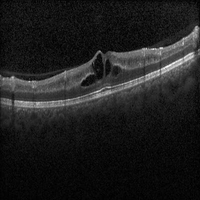
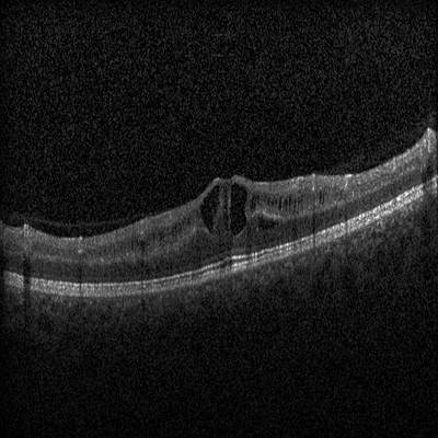
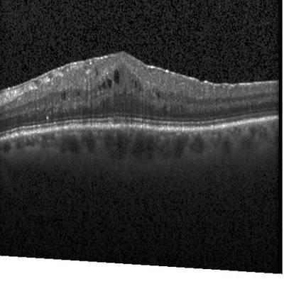
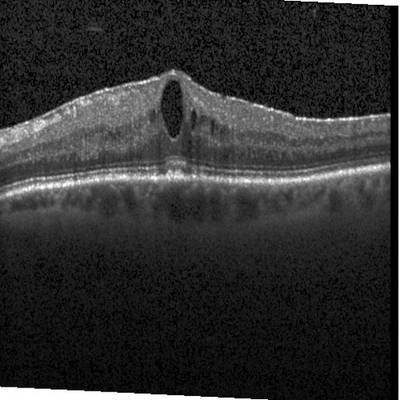
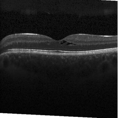
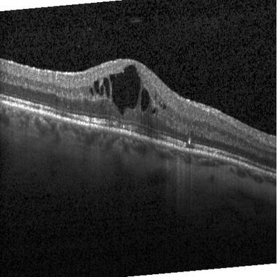
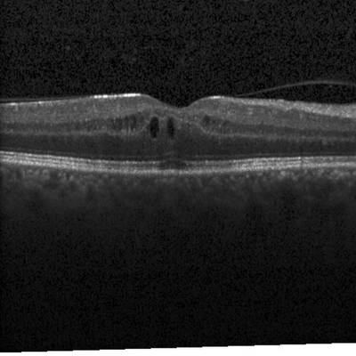
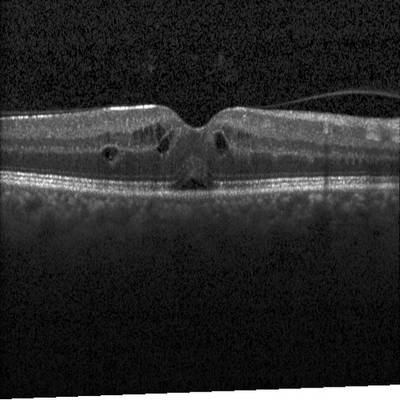
Intraretinal Cystoid Fluid dataset has 1460 images. Click on one of the examples below or open "Explore" tool anytime you need to view dataset images with annotations. This tool has extended visualization capabilities like zoom, translation, objects table, custom filters and more. Hover the mouse over the images to hide or show annotations.


Class balance #
There are 1 annotation classes in the dataset. Find the general statistics and balances for every class in the table below. Click any row to preview images that have labels of the selected class. Sort by column to find the most rare or prevalent classes.

Class ㅤ | Images ㅤ | Objects ㅤ | Count on image average | Area on image average |
|---|---|---|---|---|
cystic macular edema➔ mask | 1460 | 6542 | 4.48 | 0.55% |
Images #
Explore every single image in the dataset with respect to the number of annotations of each class it has. Click a row to preview selected image. Sort by any column to find anomalies and edge cases. Use horizontal scroll if the table has many columns for a large number of classes in the dataset.
Object distribution #
Interactive heatmap chart for every class with object distribution shows how many images are in the dataset with a certain number of objects of a specific class. Users can click cell and see the list of all corresponding images.
Class sizes #
The table below gives various size properties of objects for every class. Click a row to see the image with annotations of the selected class. Sort columns to find classes with the smallest or largest objects or understand the size differences between classes.
Class | Object count | Avg area | Max area | Min area | Min height | Min height | Max height | Max height | Avg height | Avg height | Min width | Min width | Max width | Max width |
|---|---|---|---|---|---|---|---|---|---|---|---|---|---|---|
cystic macular edema mask | 6542 | 0.12% | 4.03% | 0% | 1px | 0.2% | 182px | 35.55% | 25px | 4.99% | 1px | 0.13% | 170px | 33.2% |
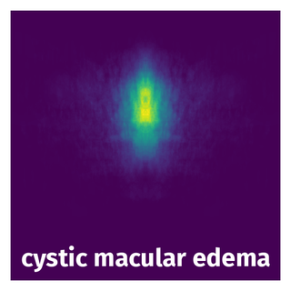
Spatial Heatmap #
The heatmaps below give the spatial distributions of all objects for every class. These visualizations provide insights into the most probable and rare object locations on the image. It helps analyze objects' placements in a dataset.
Objects #
Table contains all 6542 objects. Click a row to preview an image with annotations, and use search or pagination to navigate. Sort columns to find outliers in the dataset.
Object ID ㅤ | Class ㅤ | Image name click row to open | Image size height x width | Height ㅤ | Height ㅤ | Width ㅤ | Width ㅤ | Area ㅤ |
|---|---|---|---|---|---|---|---|---|
1➔ | cystic macular edema mask | 30DME_as_F.jpeg | 496 x 512 | 77px | 15.52% | 24px | 4.69% | 0.51% |
2➔ | cystic macular edema mask | 30DME_as_F.jpeg | 496 x 512 | 37px | 7.46% | 18px | 3.52% | 0.15% |
3➔ | cystic macular edema mask | 30DME_as_F.jpeg | 496 x 512 | 56px | 11.29% | 23px | 4.49% | 0.28% |
4➔ | cystic macular edema mask | 30DME_as_F.jpeg | 496 x 512 | 15px | 3.02% | 4px | 0.78% | 0.02% |
5➔ | cystic macular edema mask | 30DME_as_F.jpeg | 496 x 512 | 16px | 3.23% | 14px | 2.73% | 0.04% |
6➔ | cystic macular edema mask | 30DME_as_F.jpeg | 496 x 512 | 22px | 4.44% | 13px | 2.54% | 0.09% |
7➔ | cystic macular edema mask | 30DME_as_F.jpeg | 496 x 512 | 17px | 3.43% | 9px | 1.76% | 0.03% |
8➔ | cystic macular edema mask | 30DME_as_F.jpeg | 496 x 512 | 32px | 6.45% | 27px | 5.27% | 0.21% |
9➔ | cystic macular edema mask | DME155.jpeg | 512 x 512 | 13px | 2.54% | 20px | 3.91% | 0.08% |
10➔ | cystic macular edema mask | 802DME_F.jpeg | 496 x 512 | 34px | 6.85% | 17px | 3.32% | 0.14% |
License #
Intraretinal Cystoid Fluid is under CC BY-NC-SA 4.0 license.
Citation #
If you make use of the Intraretinal Cystoid Fluid data, please cite the following reference:
Zeeshan Ahmed, Munawar Ahmed, Attiya Baqai, & Fahim Aziz Umrani. (2022).
Intraretinal Cystoid Fluid [Data set].
Kaggle.
https://doi.org/10.34740/KAGGLE/DS/2277068
If you are happy with Dataset Ninja and use provided visualizations and tools in your work, please cite us:
@misc{ visualization-tools-for-intraretinal-cystoid-fluid-dataset,
  title = { Visualization Tools for Intraretinal Cystoid Fluid Dataset },
  type = { Computer Vision Tools },
  author = { Dataset Ninja },
  howpublished = { \url{ https://datasetninja.com/intraretinal-cystoid-fluid } },
  url = { https://datasetninja.com/intraretinal-cystoid-fluid },
  journal = { Dataset Ninja },
  publisher = { Dataset Ninja },
  year = { 2026 },
  month = { jul },
  note = { visited on 2026-07-26 },
}
Download #
Dataset Intraretinal Cystoid Fluid can be downloaded in Supervisely format:
As an alternative, it can be downloaded with dataset-tools package:
pip install --upgrade dataset-tools
… using following python code:
import dataset_tools as dtools

dtools.download(dataset='Intraretinal Cystoid Fluid', dst_dir='~/dataset-ninja/')
Make sure not to overlook the python code example available on the Supervisely Developer Portal. It will give you a clear idea of how to effortlessly work with the downloaded dataset.
The data in original format can be downloaded here.
Disclaimer #
Our gal from the legal dep told us we need to post this:
Dataset Ninja provides visualizations and statistics for some datasets that can be found online and can be downloaded by general audience. Dataset Ninja is not a dataset hosting platform and can only be used for informational purposes. The platform does not claim any rights for the original content, including images, videos, annotations and descriptions. Joint publishing is prohibited.
You take full responsibility when you use datasets presented at Dataset Ninja, as well as other information, including visualizations and statistics we provide. You are in charge of compliance with any dataset license and all other permissions. You are required to navigate datasets homepage and make sure that you can use it. In case of any questions, get in touch with us at hello@datasetninja.com.